Research
CNV Fabrication
Production of Cell-engineered nanovesicles (CNV)

Research into exosomes is hindered by their rarity. We tried to overcome the limitations of this exosome by developing a novel and effective nanovesicle production method.
(A)As cells flow through slits in microchannels, the cells are stretched. At the outlet of the microchannels, nanovesicles are generated possibly due to abrupt pressure change and their elongated shape caused by shear stress.
(B)After cells were loaded into the syringe, centrifugal force was applied to extrude them. During extrusion, the polycarbonate filter imposes surface tension and generates nanovesicles when they pass through the filter pores.
- Reference
- 1. Large-scale generation of cell-derived nanovesicles, Nanoscale, 2014
- 2. Mircofluidic fabrication of cell derived nanovesicles as endogenous RNA carriers, Lab on a chip, 2014
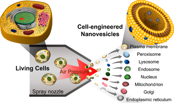
Production and characterization of Cell-engineered nanovesicles (CNV) by air spray

Living cells are inserted into the inlet and then air-sprayed; this process breaks the plasma membrane and organelle membranes into fragments, which soon spontaneously form spherical CNVs. CNVs may originate from various organelles, therefore, we clarify the origin of CNVs by measuring fluorescence expressions for organelle-specific markers using fluorescence nanoparticle tracking analysis (NTA).
- Reference
- Heterogeneous Subcellular Origin of Exosome-Mimetic Nanovesicles Engineered from Cells, ACS Biomaterials Science & Engineering, 2020